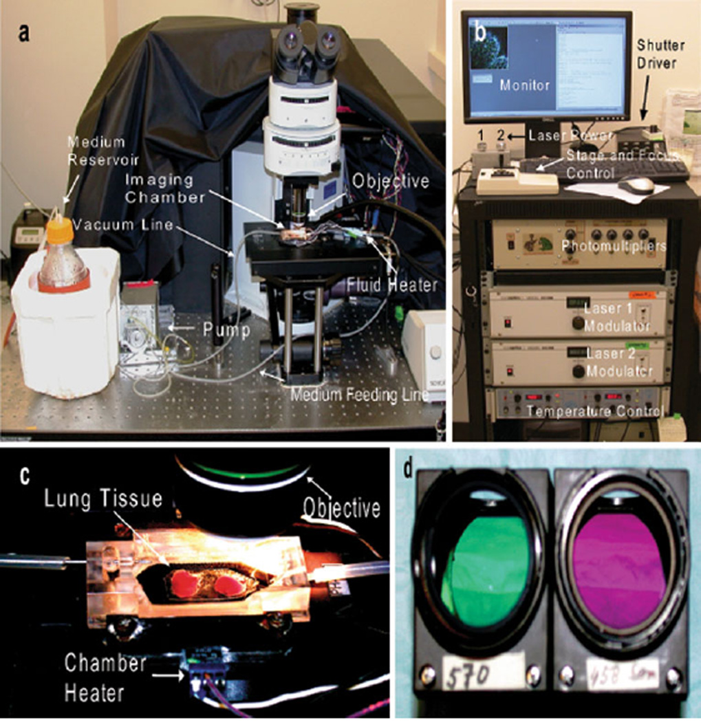
Fig. 1.
Experimental setup for imaging of lung explants with custom-built 2P microscope. a Medium is pumped from the reservoir trough a feeding line to the imaging chamber and a vacuum line evacuates the medium from the chamber. The amount of fluid flowing through the chamber is controlled by the pump. The temperatures of the chamber and fluid are maintained at 37°C throughout the experiment with separate heaters. b The acquisition console is depicted with multiple components which control the intensity of the laser as well as the movement of the stage and the focus of the objective. Images of the tissue can be observed on the monitor in real time. c Lung tissue is immersed in warm medium within the imaging chamber. d Two different filters (570 and 458 nm, respectively) housed inside dichroic cubes. Filters can be exchanged based on experimental design

