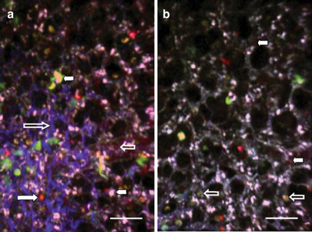
Fig. 2.
2P microscopy of lung explants. Different fluorescent probes were used for this experiment to show the versatility of the system. T cells, isolated from a wild-type C57BL/6 mouse and labeled with fluorescent dyes CMTMR or CMAC, were injected into a C57BL/6 CD11c-EYFP host. The left lung was imaged 24 h after adoptive transfer. a Quantum dots (12 µl resuspended in 150 µl of phosphate-buffered saline), injected 10 min prior to sacrifice, label blood vessels in red (small clear arrow). T cells labeled with CMTMR are observed in orange (large white arrow), and T lymphocytes labeled with CMAC are seen in cyanide (white arrowhead). Host CD11c+ cells with dendritic morphology appear yellow-green (small white arrow). SHG depicts collagen in blue, which can be seen delineating air spaces (large clear arrow). b Alveolar macrophages display characteristic nuclear shadow (clear arrows). Peri-alveolar blood vessels have been labeled with quantum dots and appear red (white arrows). Space bar represents 30 µm

