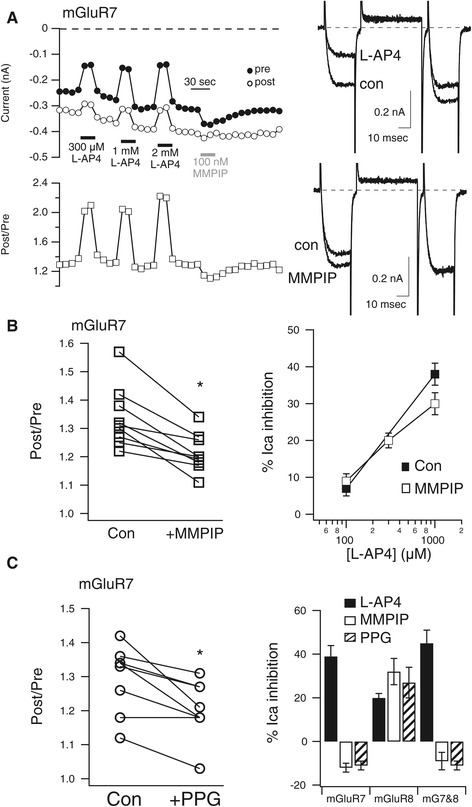
Figure 3.

mGluR7 is a constitutively active receptor. A, Time course of calcium current amplitudes (upper left) and facilitation ratios (lower left) from the “pre” and “post” test pulses obtained with the triple pulse protocol in a representative SCG neuron during application of the indicated concentrations of L-AP4 and 100 nM MMPIP. Sample current traces from the same cell are shown to the right. Control and L-AP4 inhibited currents are shown (upper right), as are control and MMPIP enhanced currents (lower right). B, Plot of control facilitation values and those in the presence of MMPIP (left) in mGluR7 expressing neurons, paired from each cell. * indicates significant difference (p < 0.05, paired T-test). L-AP4 concentration-response curve is also shown for two [L-AP4]s when applied alone (■), and three [L-AP4]s when applied in the presence of 100 nM MMPIP (□). No significant differences were detected. C, Plot of control facilitation values and those in the presence of PPG (left) in mGluR7 expressing neurons, paired from each cell. *indicates significant difference (p < 0.05, paired T-test). Average responses (±SEM) to L-AP4 (black bars), MMPIP (white bars), and PPG (striped bars) in SCG neurons expressing mGluR7, mGluR8, and mGluR7&8 together, as indicated.
