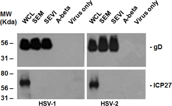
Figure 1.
Amyloid-virus binding assay. (Left) 1 mL of HSV-1 (107/mL) was incubated with 5 μg of SEVI or SEM amyloid fibrils for 1 h at 37 °C. Controls include virus alone and virus incubated with 5 μg of A-beta amyloid. The mixtures were centrifuged and the pellet was washed twice with MEM. The pellet was lysed in 2× Laemmli buffer and blotted using anti-ICP27 (non-structural protein) and anti-gD (envelope protein) antibodies. Whole cell lysates (WCL) of HSV-1–infected 293T cells were used as input controls for the Western blot; (Right) Similar conditions, but using HSV-2 instead of HSV-1. WCL = whole cell lysate made from HSV-infected HEK 293T cells.

