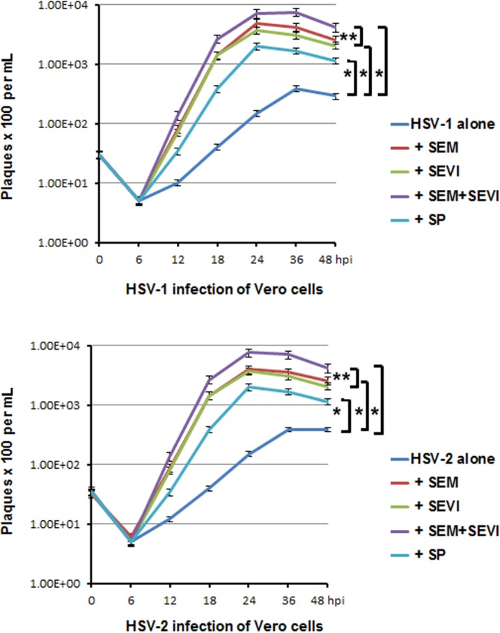
Figure 5.
Viral growth curve assay. (Upper) HSV-1 infection of Vero cells. Vero cells were infected with virus alone or with virus treated with SP (1:1000 dilution), SEM amyloids (5 μg/mL), SEVI (5 μg/mL), or SEM amyloids (5 μg/mL) plus SEVI (5 μg/mL) at an MOI of 0.1 for a differing number of hours (as indicated). One well of each culture was collected (along with medium) at the indicated hpi and stored at −80 °C. The collected cells (with medium) were frozen/thawed for three cycles and then centrifuged at 8000 g for 20 min to remove the cellular debris. To perform a pfu assay, 25 μL of supernatant of each sample was used to infect Vero cells in triplicate; (Lower) the same as described in the above, except that the Vero cells were infected with HSV-2. Student’s t-test was used to statistically analyze the differences between the groups versus virus alone (*p < 0.001) and those between the group of SEM + SEVI versus SEM or SEVI (**p < 0.005).

