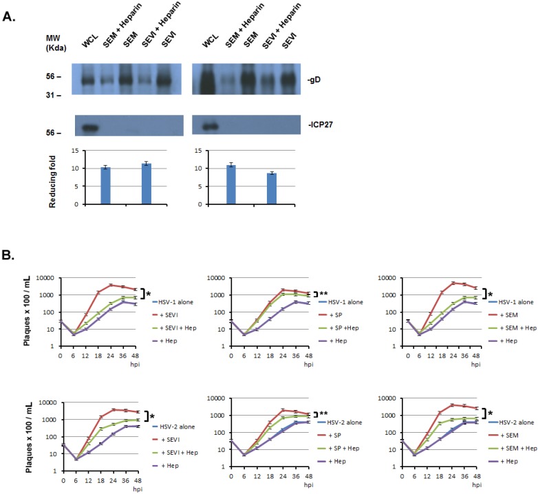
Figure 6.
Heparin inhibits the amyloids’ enhancement of HSV infection. (A) Heparin interferes with the binding of HSV with amyloids. One milliliter of HSV-1 (107/mL) was incubated with 5 μg of SEVI or SEM amyloid fibrils in the absence or presence of heparin (5 μg/mL) for 1 h at 37 °C. Controls included virus alone and virus incubated with 5 g of A-beta amyloid. The mixtures were centrifuged and the pellet was washed twice with MEM. The pellet was lysed in 2× Laemmli buffer and blotted using anti-ICP27 (non-structural protein) and anti-gD (envelope protein) antibodies. Whole cell lysates (WCL) of HSV-infected 293T cells were used as input controls for the Western blot. The density of the gD bands of HSV-1 or HSV-2 from non-heparin treated was compared with that of treated heparin, the fold increase of gD levels for HSV-1 (left) and for HSV-2 (right) were calculated (Quantity One software, version 4.5.0, Bio-Rad Laboratories, Richmond, CA, USA) and are shown below the corresponding Western blots; (B) Heparin significantly reduced the enhancing effects of SEM or SEVI on HSV infection. PFU assays were conducted in Vero cells using the same protocol as that described in Figure 5, except that heparin (5 μg/mL) was used in these experiments. Student’s t-test was used to statistically analyze the differences between the group of virus treated with amyloid (SEM or SEVI) versus those between virus treated with amyloids plus heparin (Hep) (*p < 0.001) or the differences between the group of virus treated with SP versus those between virus treated with SP plus heparin (Hep) (**p < 0.05).

