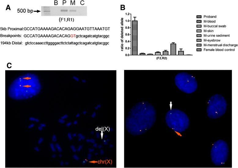
Figure 3.

Varying proportions of a mosaic deletion among different tissue samples from the mother. (A) Breakpoint analysis for the deletion in the proband and his mother. Upper panel: Electrophoresis results for the amplified products with primers F1 and R1, which show bands of about 550 bp for both the proband and his mother. This amplification failed for normal controls due to the huge span, suggesting a same deletion in the mother and in her son. B: blank control, P: proband, M: mother, C: normal female control. Bottom panel: Sequence analyses of the breakpoints in the proband and the mother revealed an insertion (red) of two nucleotides (GT) at the junction. This deletion extended approximately 5 kb proximal to and 194 kb distal of the FMR1. (B) qPCR analyses of the deleted alleles show varying proportions of mosaicism among multiple tissues from the mother, including her eyebrow, buccal swab, skin, urine sediment and menstrual discharge. The mosaic proportion of the deleted alleles was low in the blood (4%), while it was higher in the skin (8%), urine sediment (11%), menstrual discharge (12%) and eyebrow (33%). Error bars indicate standard deviations. (C) Representative FISH analysis of the mother’s skin-derived fibroblasts. Left panel shows an abnormal mitosis with one X chromosome that has only the green control signal (indicated by the white arrow), suggesting a loss of the complementary fragment in this cell. Neighbouring nuclei had both targeted and control signals on both X chromosomes, suggesting a normal genotype. Right panel shows an abnormal nucleus (indicated by the white arrow) surrounded by four nuclei with normal genotype. A total of 213 abnormal cells (9 mitoses and 204 nuclei) with single targeted signal and 2 control signals were detected based in 1600 counts, which indicated mosaicism of 13% in the mother’s fibroblasts.
