
Figure 1.
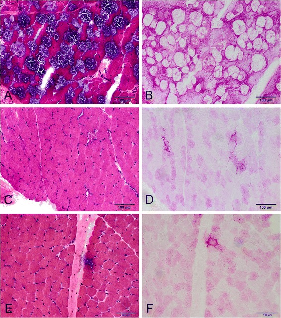
Myopathological changes in Patient 2 (A and B), Patient 12 (C and D) and Patient 22 (E and F). H&E staining shows extensive vacuolation in many fibers in Patient 2 (A), but only a few vacuolar fibers in Patient 12 (C) and Patient 22 (E). Vacuolar fibers stained positive for glycogen with PAS (B, D and F).