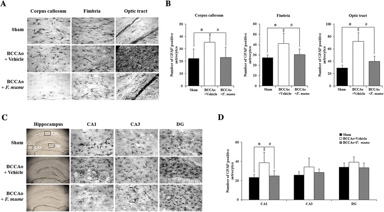
Figure 3.

Effects of F. mume on chronic BCCAo-induced astrocytic activation in the white matter. Immunohistological staining was conducted to investigate the expression of GFAP-positive cells in the white matter and the hippocampus in the sham-operated group (n = 7-8), the BCCAo + Vehicle group (n = 9), and the BCCAo + F. mume group (n = 9). Representative photomicrograph of GFAP (A and B) positive cells. The number of GFAP (C and D)-positive cells was increased in the cerebral cortex, fimbria, the optic tract and CA1 of the BCCAo-injured rats compared to the sham-operated rats (*), and was significantly decreased in the optic tract of the BCCAo rats treated with F. mume compared to the BCCAo-injured rats (#). Data were analyzed via ANOVA followed by the Tukey test. *, p < 0.05 versus the BCCAo + Vehicle group; #, p < 0.05 versus the BCCAo + F. mume group. CA 1 and 3, cornuammonis 1 and 3; DG, dentate gyrus.
