
Figure 4.
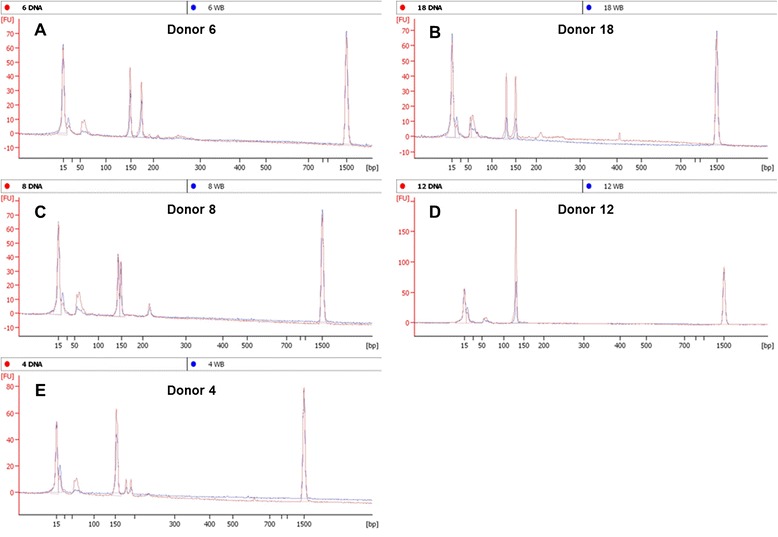
Selected bioanalyzer electropherogram overlays from purified DNA (red) or 10% whole blood (blue). Results from five representative donors illustrate examples of a confirmed heterozyote (A), heterozygote with a 10% blood under the default FU cutoff (B), and suspected heterozygote with split peaks as an allelic differentiator and a larger artifact (C), confirmed homozygote (D), homozygote with two smaller artifacts (E).