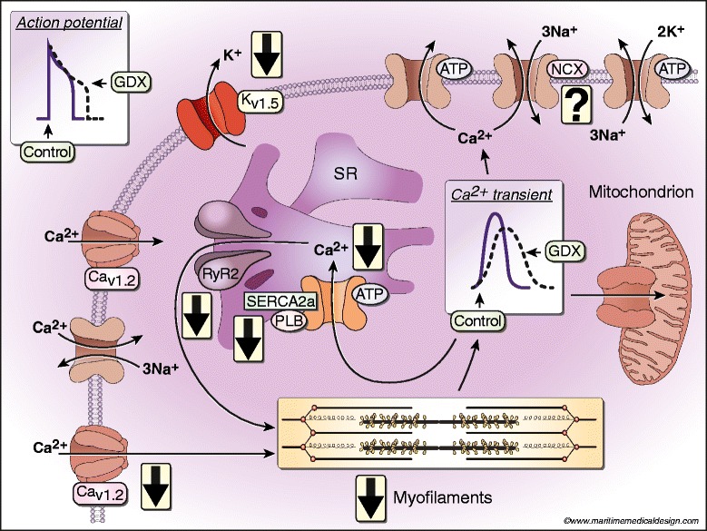
Figure 2.

Impact of GDX on intracellular Ca2+-handling mechanisms in ventricular myocytes isolated from rodent hearts. APD is prolonged by GDX, due to a decrease in repolarizing K+ currents (IKur) and a reduction in the expression of Kv1.5. Reduced Ca2+ influx along with smaller Ca2+ sparks attenuates SR Ca2+ release. Ca2+ transient decay is slowed by longer APs and slower SR Ca2+ uptake mediated by a decrease in phosphorylation of PLB by CaMKII (and possibly PKA). Peak contractions are attenuated through smaller peak Ca2+ transients and a decrease in maximal myofilament responsiveness to Ca2+. Contractions are slowed because SR Ca2+ uptake is reduced and the slower β-MHC isoform predominates. Whether NCX activity or expression is affected by GDX is not yet clear.
