
Figure 6. DRN Pet-1 neurons release 5-HT and glutamate.
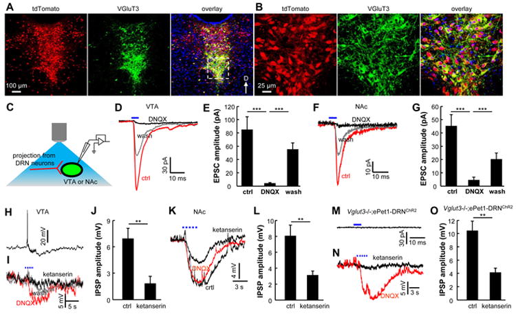
(A and B) In an ePet1-Cre;Ai14 mouse, VGluT3 (green) is expressed in many tdTomato-labeled neurons (red) along the midline. Panels in (B) show the zoom-in view of the dashed rectangular area in (A). (C) Schematic diagram showing the method of optogenetic stimulation and recordings from the VTA or the NAc in brain slices. (D and E) Representative recording traces from a VTA neuron (D) and group data (E) reveal that brief light stimulation of ChR2+ axonal terminals produced fast EPSCs that were reversibly blocked by DNQX (***, p<0.001; paired t-tests; n = 13 cells). (F and G) Glutamatergic EPSCs were also evoked by single-pulse light stimulations in the NAc shell (***, p<0.001; paired t-tests; n=7 cells). (H and I) Current-clamp recordings from a single VTA neuron show that trains of light pulses (3 s, 20 Hz) resulted in brief excitation, followed by slow inhibition (H). The initial excitatory response was blocked DNQX, whereas the slow inhibitory response was largely abolished by ketanserin, which blocks 5-HT2A and 5-HT2C receptors (I). (J) Group data showing the effect of ketanserin on the slow IPSPs (**, p<0.01; paired t-test; n = 6 cells). (K and L) Slow 5-HT effects were also observed in the NAc (**, p<0.01; paired t-test; n = 7 cells). (M and N) Brief light stimulation failed to elicit any fast EPSC from a cell in the VTA of a Vglut3-/-;ePet1-DRNChR2 mouse (M), but repetitive light stimulation (3 s, 20 Hz) evoked slow IPSP that was largely abolished by ketanserin (N). (O) Group data showing that the slow IPSPs were significantly reduced by ketanserin in Vglut3-/-;ePet1-DRNChR2 mice (*, p<0.01; paired t-test; n = 6 cells). See also Figure S6.