
Figure 7. Data from iClass tests and two-bottle preference tests reveal that both 5-HT and glutamate contribute to reward signaling by DRN Pet-1 neurons.
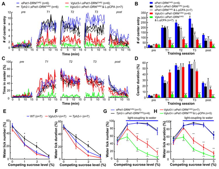
(A and B) In iClass tests, Tph2-/-;ePet1-DRNChR2 mice and L-pCPA-treated ePet1-DRNChR2 mice showed a mild but statistically significant reduction in the center entry number for certain training sessions (T2 or T3). Vglut3-/-;ePet1-DRNChR2 exhibited ∼50% reduction in the number of center entries of all training sessions. L-pCPA injection into Vglut3-/-;ePet1-DRNChR2 mice completely abolished the reward effect produced by the activation of DRN Pet-1 neurons.*, p<0.01; ***, p<0.001; t-tests between test groups and ePet1-DRNChR2 control mice. (C and D) The effect of knocking out the Vglut3 gene and/or depleting 5-HT on the center duration. (E and F) The sucrose preference scores quantified with lick numbers and lick duration, respectively. Both Tph2-/- and Vglut3-/- mice preferred sucrose to water, but the sucrose preference scores of Tph2-/- mice were lower than those of wild-type mice at the concentrations of 1 and 2%. *, p < 0.05; Two-way ANOVA and then Dunnett's multiple comparison tests between mutants and WT. (G and H) Sucrose preference scores show that light stimulation of the DRN Pet-1 neurons in Vglut3-/-;ePet1-DRNChR2 mice produced a reward value of ∼1% sucrose. L-pCPA injection into these mice completely disrupted reward signaling. **, p<0.01; ***, p<0.001; one-way ANOVA and then Tukey's post-hoc test between test groups and ePet1-DRNChR2 control mice. See also Figure S7.