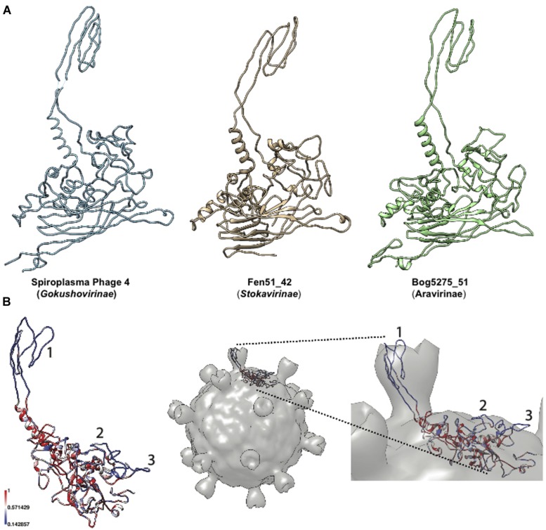
FIGURE 2.
Modeling of major capsid proteins. (A) Three-dimensional models of Microviridae major capsid protein. Different subfamilies are depicted with, from left to right, the reference model from Spiroplasma Phage 4 (Pdb Id: 1KVP), representatives from Stokavirinae and Aravirinae. (B) Hypervariable regions of Aravirinae major capsid protein. The sequence conservation across the seven Aravirinae sequences was mapped on the three-dimensional model obtained for the Bog5275_51 genome. The three hypervariable regions were numbered (left panel). This model was mapped on the capsid structure of Spiroplasma phage 4 to estimate the position of these hypervariable regions relative to the whole virion (right panel).

