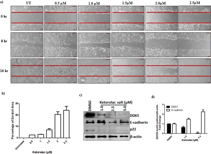
Figure 4. Ketorolac salt inhibits the growth of human OSCC: a) H357 cells were placed on 6 well culture plates.
After the cells became confluent (monolayer) scratch was made in each well with the help of a 10 μL micropipette tip to generate a uniform wound that was devoid of adherent cells. Wells were treated with DMSO and with indicated concentration of Ketorolac salt. The photograph was taken using inverted microscope b) Bar graph showing quantification of the scratch area. c) H357 cells were treated with indicated amount of Ketorolac salt for 48h and lysate was prepared, western blotting was performed to detect the expression of DDX3, E-cadherin, p21 and β-actin. d) The Bar graph indicates the ratio of DDX3 and E-cadherin to β-actin band intensity. SD (n = 3).

