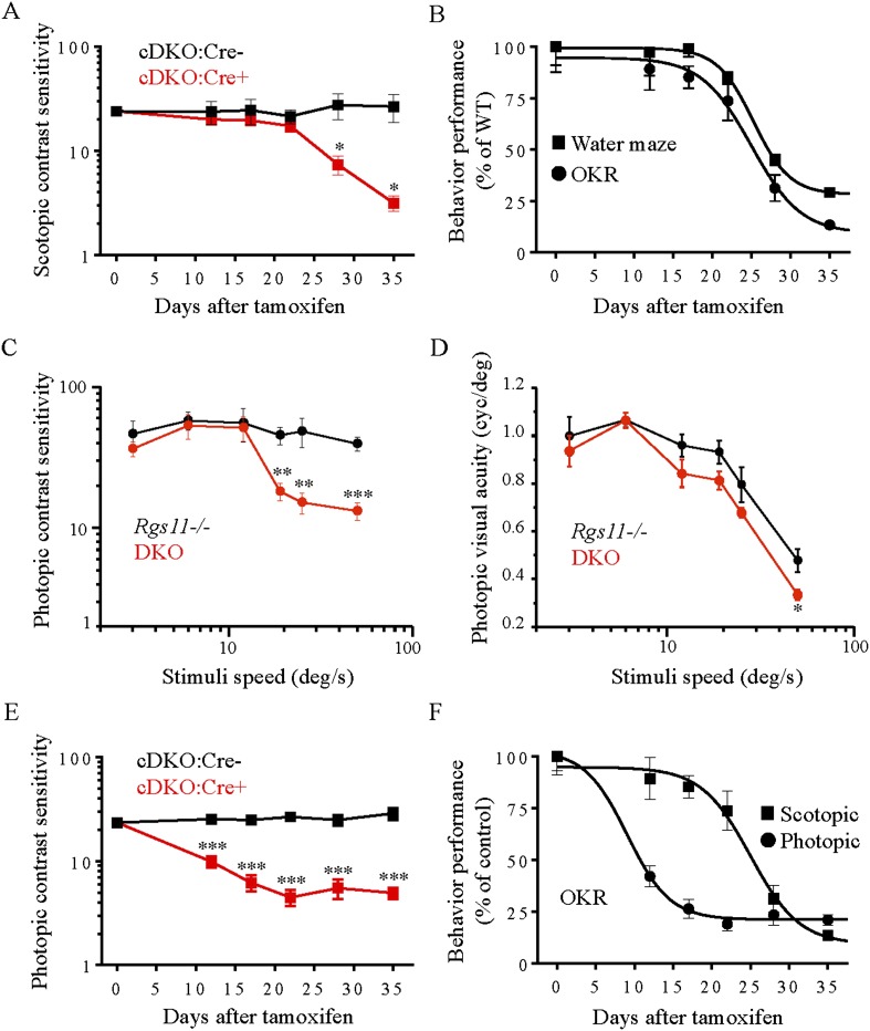
Figure 9. Evaluation of scotopic and photopic mouse vision by optomotor task.
(A) Scotopic (∼3 × 10⁻⁴ cd/m2) contrast sensitivity deficits in mice lacking RGS proteins (cDKO:Cre+) vs cDKO:Cre- mice as a function of time of post-tamoxifen administration (*p < 0.05, t-test, n = 4). (B) Comparison of visual performance of mice in water maze task and optomotor response tests. Mouse vision declined by half after 25 ± 1 days of tamoxifen administration in both experiments. (C) The reliance of high speed contrast sensitivity on intact ON-BC function. Deficits in photopic (70 cd/m2) contrast sensitivity of DKO mice are apparent at stimuli speeds of >12 deg/s (error bars are SEM; t-test: **p < 0.01, ***p < 0.001, n = 4–5). (D) Visual acuity is mainly unaffected by the loss of RGS11 together with RGS7 (DKO) as compared to Rgs11−/− controls. (error bars are SEM. t-test: *p < 0.05, n = 4–5 mice). (E) Photopic (70 cd/m2) contrast sensitivity deficits in mice lacking RGS proteins (cDKO:Cre+) vs cDKO:Cre- controls as a function of time of post-tamoxifen administration (error bars are SEM; t-test: ***p < 0.001, n = 7). (F) Comparison of progressive RGS elimination effects on scotopic vs photopic contrast sensitivities of mice. The half-time of contrast sensitivity decline is 25 ± 1 days for scotopic vs 9.0 ± 2 days for photopic conditions, respectively. The data for each group are normalized to their respective values upon beginning of the tamoxifen treatment (day 0) and expressed as percentage.

