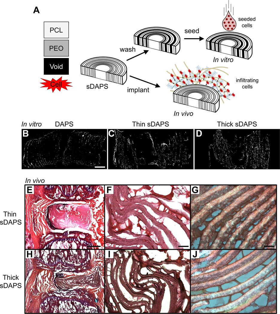
Figure 7. Sacrificial DAPS (sDAPS) improve cell colonization in vitro and in vivo.
(A) To overcome issues of poor cell infiltration, sDAPS were fabricated with a layer of PEO spun directly onto a PCL layer prior to wrapping. sDAPS were evaluated both in vitro (via surface seeding of constructs with bovine AF cells and 7 days of culture) and in vivo (via direct implantation). DAPI staining of cross sections on day 7 show increasing cell infiltration in vitro comparing PCL-only DAPS (B), ‘thin’ sacrificial layer sDAPS (C), and ‘thick’ sacrificial layer sDAPS (D). Implantation in the rat caudal spine (with external fixation) showed improved colonization and matrix deposition in ‘thin’ sDAPS (E–G) and ‘thick’ sDAPS (H–J). Matrix formation between lamellae was apparent at higher magnification in both formulations (F, G and I, J). Scale = 500 µm in (B), 1 mm in panel (E), 100 µm in panel (F), and 50 µm in panel (G).

