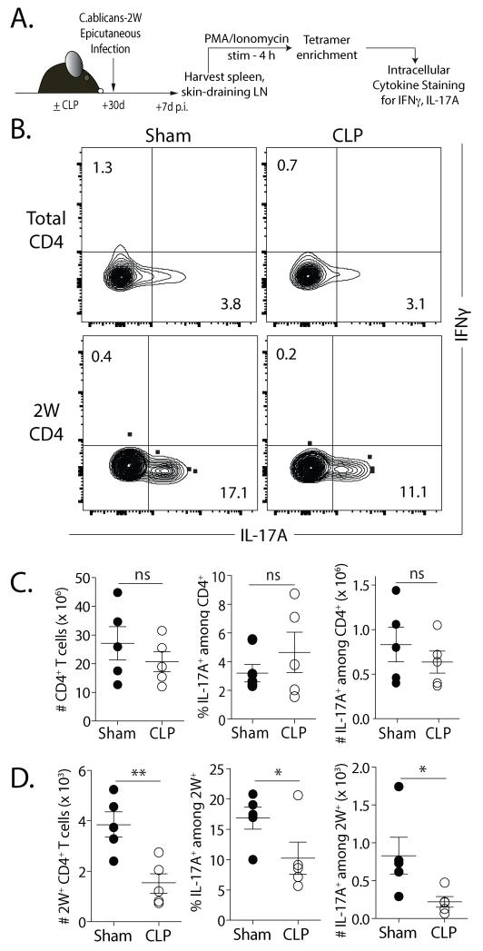
FIGURE 6. Ag-specific CD4 T cells have functional deficits in Th17-polarized responses.
A. Experimental design. On d 30 after sham or CLP surgery, mice were infected epicutaneously with 2W1S-expressing C. albicans (C. albicans-2W; 108 yeasts in 0.05 ml). After 7 d, lymphocytes obtained from the skin-draining (inguinal, brachial, axillary and cervical) LN of infected mice were stimulated for 4 h with PMA/ionomycin. The stimulated samples were enriched for 2W1S-specific CD4 T cells and production of IFNγ and IL-17A was assayed by flow cytometry. B. Representative flow plots showing the gating strategy to identify IL-17A+IFNγ− cells within bulk and 2W:I-Ab-specific CD4 T cells. C-D. Frequency and number of IL-17A+ in bulk (C) and 2W1S-specific CD4 T cells (D) in infected sham- or CLP-treated mice. Statistical significance was determined using Welch”s t-test. ** p < 0.01; * p < 0.05; and n.s. – not significant. Data shown are representative results from 2 independent experiments, with 5 mice/group in each experiment.

