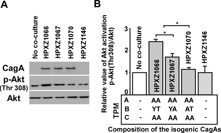
Figure 3. Analysis of the PI3-kinase-AKT pathway after co-culture of human AGS cells with isogenic H. pylori strains containing the engineered CagA molecules.

Panel A: After 24 h co-culture with H. pylori isogenic cagA mutants, AGS cells were washed 5 times with ice-cold PBS buffer to remove H. pylori cells, and whole cell lysates were separated by SDS-PAGE, followed by immunodetection with antibodies (α-CagA, α-p-AKT, or α-AKT). Panel B: Targeted bands were quantified with ImageJ software to calculate the relative level of AKT phosphorylation.
