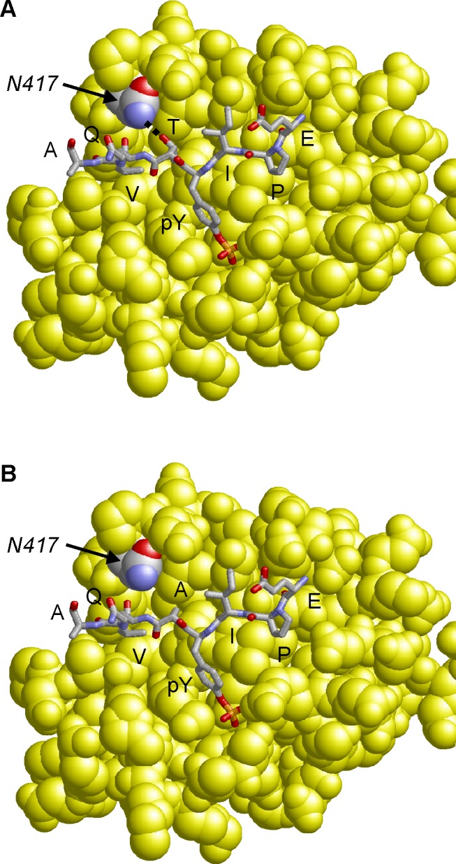
Figure 6. Model of the CagA B-TPM motif variants bound to PI3-kinase.

The interactions of the EPIYTQVA motif (Panel A) are compared to that of the EPIYAQVA motif (Panel B). The threonine residue at the pY+1 position forms a side-chain hydrogen bond to N-417 of PI3-kinase, which cannot be formed by alanine. The motif is shown in stick presentation and colored according to atom type. The PI3-kinase SH2-domain is shown in yellow space-filled presentation and N-417 is colored by atom type.
