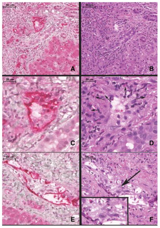
Figure 2.

Composite histopathological features of severe, acute, C4d+ antibody mediated rejection (AMR) (A-F). A) Note intense and diffuse C4d staining (red) in the portal vein (PV) and portal capillaries (*). B) Routine H&E appearance of the same portal tract as shown in A). Note the marked endothelial cell hypertrophy of the portal venous endothelium (*). C) Shows a C4d stain (red) of the same portal vein (PV) branch as B) at higher magnification. D) Shows the routine H&E appearance of this vein. Note the marked portal venous endothelial cell hypertrophy intermixed with eosinophils and histiocytes. E) Another C4d staining example of the portal venous changes typical of severe acute AMR with the H&E counterpart shown in figure F). G) In contrast, C4d-negative T-cell mediated rejection (control case) shows predominantly mononuclear portal inflammation with focal subendothelial localization of the lymphocytes in a small portal vein branch (arrow). H) Routine H&E appearance of a control case's portal infiltrate highlights several differences: the substantial decrement in edema, the absence of marked portal microvascular endothelial cell hypertrophy, and the marked diminution in eosinophils. A scale bar is shown at the top left of each image.
