Fig. 1.
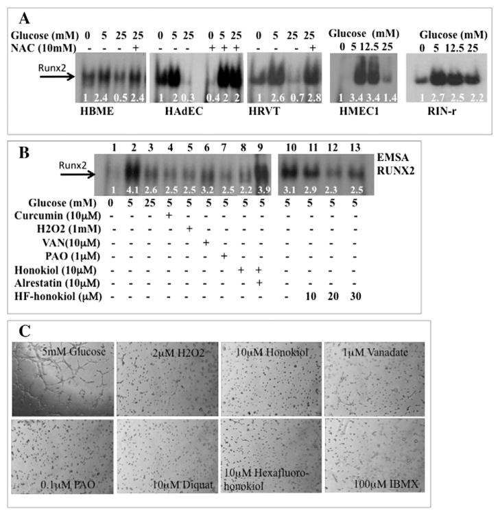
Regulation of RUNX2 DNA binding by HG and oxidative stress. (A) Human bone marrow microvascular ECs (HBME), human adipose ECs (HAdEC), human microvascular dermal ECs (HMEC1), telomerized human umbilical vein ECs (HRVT), or rat pancreatic β-cells (RIN-r) were cultured in starvation media for 16 h and treated with 0, 5, 12.5, or 25 mM glucose for 4 h or with 10 mM of the anti-oxidant N-acetyl Cys (NAC). Nuclear extracts were prepared and RUNX2 DNA binding activity measured by EMSA with a RUNX2-specific oligonucleotide. Assays were repeated three times. Relative band intensity is shown for a representative set of results, normalized to total protein in each lane. (B) Glucose and serum-starved ECs were treated with 5 mM or 25 mM glucose or with 5 mM glucose and the oxidants curcumin, H2O2 honokiol, HF-honokiol or the phosphatase inhibitors VAN or PAO before isolation of nuclear extracts. Relative band intensities are indicated. (C) Oxidants inhibit EC tube formation. ECs were treated with 5 mM glucose or glucose + oxidants (H2O2, diquat, honokiol, hexafluoro-honokiol, IBMX) or phosphatase inhibitors (PAO, orthovanadate), for 6 h on matrigel. Essentially similar results were obtained in three other experiments.

