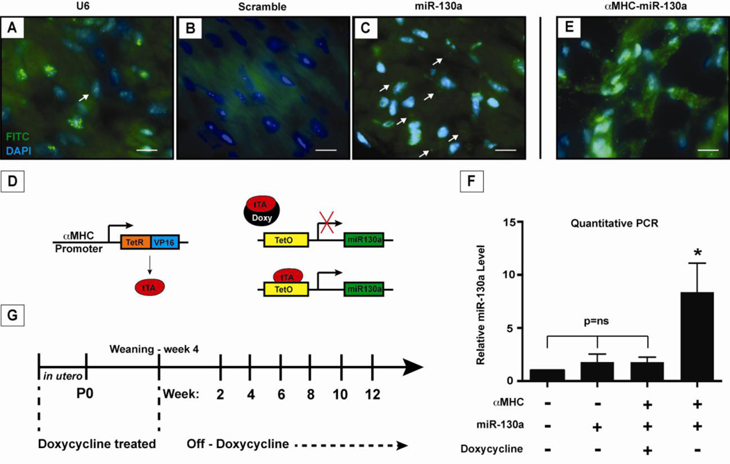
Figure 1. Expression of miR-130a in cardiomyocytes and induction of miR-130a in adult heart.
Localization of miR-130a using fluorescent in situ hybridization in adult cardiomyocytes. Panel (A) shows U6 stained nuclei as a positive control, panel (B) shows staining with a miR-scramble as a negative control, and panel (C) with miR-130a staining in cardiomyocytes. (D) Schematic of inducible transgenic system for miR-130a overexpression using the αMHC promoter to drive expression of the tTA protein. In panel (E), fluorescent in situ hybridization of αMHC-miR130a heart demonstrating increased miR-130a signal confirming overexpression of miR-130a. (F) Quantitative PCR of relative miR130a levels in transgenic heart 6–8 weeks after doxycycline is removed from water supply (n=5). (G) Schematic outline of doxycycline treatment and study time points. * indicates p<0.0001. Doxy: doxycycline; TetO: tetracycline responsive promoter; tTA: tetracycline-controlled transactivator.

