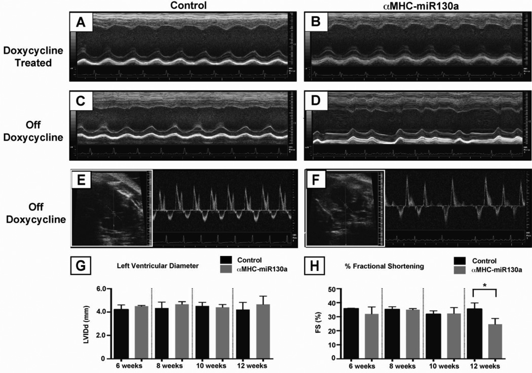
Figure 2. Echocardiography in αMHC-miR130a mice.
Panels (A) and (B), m-mode echocardiography of representative control vs. αMHC-miR130a mouse maintained on doxycycline at 10 weeks after weaning. Cardiac dimensions and function was preserved. In panels (C) and (D), representative M-mode echocardiography of control vs. αMHC-miR130a mouse at 10 weeks after doxycycline removal. Note the irregularity of ventricular contractions in the αMHC-miR130a mice. In panels (E) and (F), 2D–guided pulsed Doppler (see inset) of the mitral inflow in control and the αMHC-miR130a mice respectively. (G) and (H), serial assessment in LV diameter and % fractional shortening at 6, 8, 10, and 12 weeks. In each group, n = 5. * indicates p < 0.001.

