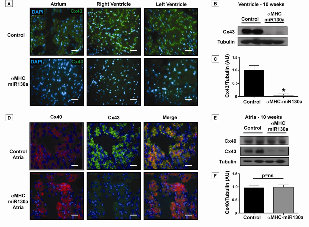
Figure 6. Loss of connexin43 in the atria and ventricles of αMHC-miR130a mice.
In panel (A), representative immunofluorescent staining of Cx43 (green) in adult control atria and ventricles compared to αMHC-miR130a atria and ventricles (n=16 hearts studied). Cx43 is reduced throughout the myocardium of αMHC-miR130a mice. (B) Western blot of ventricular lysate at 10 weeks off doxycycline demonstrating >90% reduction in Cx43 levels as quantified in (C) (n=6 per group). In panel (D), representative immunofluorescent staining of atrial tissue for Cx40 (red) and Cx43 (green) at 10 weeks off doxycycline. Note the loss of Cx43 in αMHC-miR130a atria with preserved Cx40 expression. In panel (E), atrial lysates were used for western blotting confirming preserved Cx40 levels in αMHC-miR130a atria compared to controls and quantified in (F) (n=4 per group). Consistent with the ventricular lysates, Cx43 levels were significantly reduced in the atrial lysates. Nuclei are counterstained with DAPI (blue). * indicates p < 0.001.

