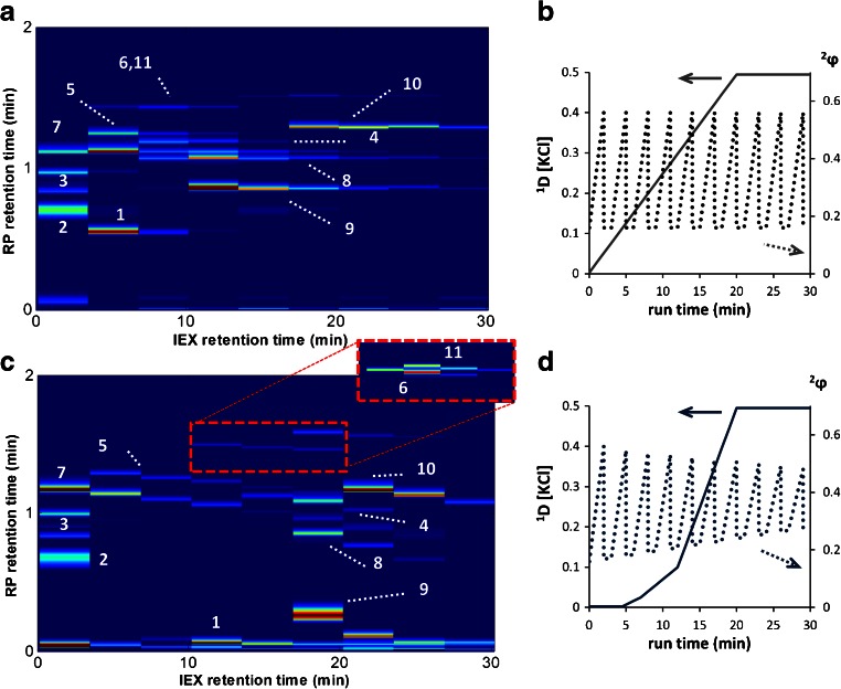
Fig. 6.
A Separation of 11 proteins by LC×LC-UV using the method as shown on the right. B Gradient conditions for separation shown in A on the left axis the concentration of counter ion and on the right axis 2 φ. C Separation of the same protein mixture as in A using the optimized method D with zoom-in illustrating the baseline separation of proteins 6 and 11. D Gradient conditions for separation shown in C. Proteins eluted were 1 ribonuclease A, 2 ovalbumin, 3 carbonic anhydrase, 4 transferrin, 5 α-lactalbumin, 6 β-lactoglobulin B, 7 trypsinogen, 8 lysozyme, 9 cytochrome c, 10 myoglobin, 11 β-lactoglobulin A

