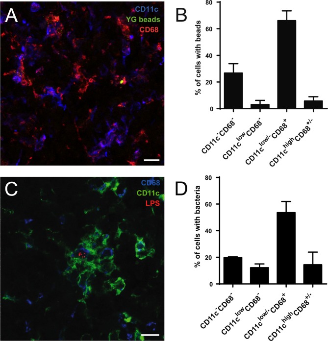
Fig 5. Brucella and fluorescent microspheres in the CLN localize in cells positive for CD68 and low or negative for CD11c.
(A) C57BL/6 mice were fed by oral gavage with 0.2 μm yellow green fluorescent microspheres. After 3 days, they were sacrificed and CLN processed for immunofluorescence microscopy. (B) Cells with internal beads from experiments as shown in (A) were quantified as to their expression of CD11c and CD68. At least 100 bead-containing cells per experiment were counted. (C) Mice infected by the oral route with 109 B. melitensis per mouse were sacrificed at day 8, CLN prepared for immunofluorescence analysis as described above and (D) the number of infected cells positive for either marker was determined. All available cuts from the CLN of one mouse were analyzed. Data shown represents mean and standard deviation of three independent experiments. Bars: 10 μm.

