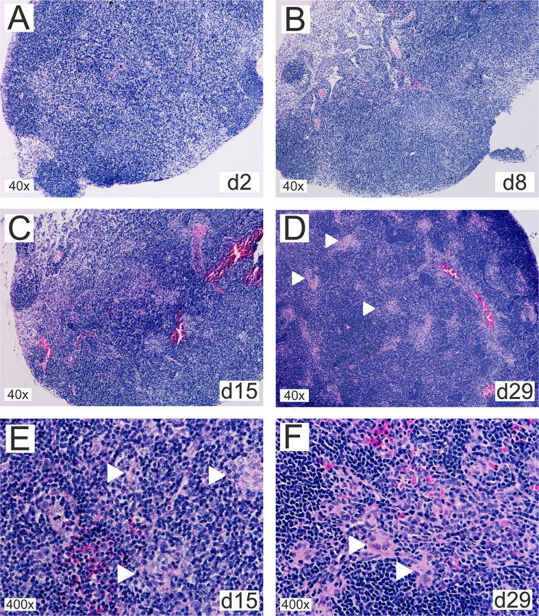
Fig 9. Oral infection with B. melitensis results in CLN granuloma formation.
Thin sections of cervical lymph nodes from mice orally infected with 109 B. melitensis per mouse for (A) 2, (B) 8, (C) 15, or (D) 29 days were stained with eosin-hematoxylin. (E) and (F) show higher magnifications from day 15 and 29, respectively. White arrowheads mark granulomatous structures that (E) develop as multifocal loose cell arrangements that (F) gradually solidify into compact granulomas composed of epithelioid cells with occasional multinucleated giant cells and few neutrophils.

