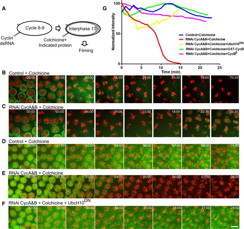
Figure 2. Full Stabilization of Cyclin B3 Requires Both the SAC and Early Cyclins.
(A) Schematic of the experiments showing the approximate stage of each injection.
(B and C) Colchicine injection induced mitotic arrest in control, but not in Cyclin A+B knockdown, embryos. Mitotic chromosomes were visualized by H2AvD-RFP (red), and nuclear envelope was visualized by injection of fluorescently labeled wheat germ agglutinin (WGA) (green). Compare based on indicated timing (min:s) not alignment.
(D–F) Characterization of Cyclin B3 degradation under different conditions. Cyclin B3-GFP (green) was stabilized by colchicine in wild-type, but not in Cyclin A+B-depleted, embryos. APC/C inhibitor UbcH10C114S blocked this destruction. Mitotic chromosomes were visualized by H2AvD-RFP (red). Note that in (E) chromosomes exited from mitosis after Cyclin B3-GFP degradation. Scale bar, 5 mμ.
(G) Quantitation of Cyclin B3-GFP fluorescent intensity after colchicine injection. Total fluorescence of each frame was measured, normalized, and plotted against time (minutes into mitosis). Cyclin B3-GFP was similarly stabilized by injection of recombinant Cyclin B proteins and a known inhibitor of the APC/C, UbcH10C114S.
See also Figures S1 and S2 and Movie S2.

