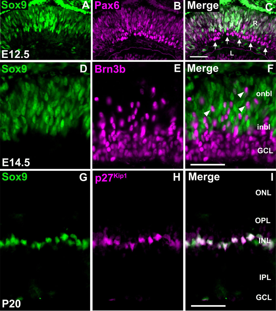
Fig. 3.
Sox9 is not expressed in retinal ganglion cells, while expression in postnatal Müller glial cells persists. E12.5 retinae were colabeled with antibodies against Sox9 and Pax6 (A–C). Clear colo-calization is observed within the retinal ventricular zone. Within the developing ganglion cell layer, which consists of Pax6 + differentiating retinal neurons, Sox9 is not expressed (arrow in C). Colabeling for Sox9 and Brn3b (D–F) confirms the above-mentioned results as Brn3b + ganglion cells do not express Sox9. These cells appear to be traversing the Sox9+ ventricular zone toward the developing GCL (arrowheads in F). At P20, Sox9 protein expression is confined to the inner nuclear layer (G). Colabeling with Sox9 and p27Kip1 revealed that postnatal retinal expression of Sox9 is specific to Müller glial cells (G–I). R, retina; L, lens; onbl, outer neuroblastic layer; inbl, inner neuroblastic layer; ONL, outer nuclear layer, INL, inner nuclear layer; GCL, ganglion cell layer; OPL, outer plexiform layer; IPL, inner plexiform layer. N = 3 mice per timepoint. Scale bars = 50 µm.

