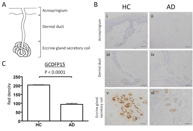
Fig 2. Immunohistochemical staining for GCDFP15 in eccrine sweat glands of normal and atopic dermatitis skin specimens.
(A) Eccrine sweat glands are composed of eccrine gland secretory coil, dermal duct, and acrosyringium. (B) Normal and AD skin specimens were immunohistochemically stained with anti-GCDFP15 antibody. Bars indicate 50 μm. (C) The staining intensities are expressed as red density (RD). The RD of GCDFP15 in the eccrine gland secretory coil is significantly lower in AD patients. Results are expressed as mean ± SEM.

