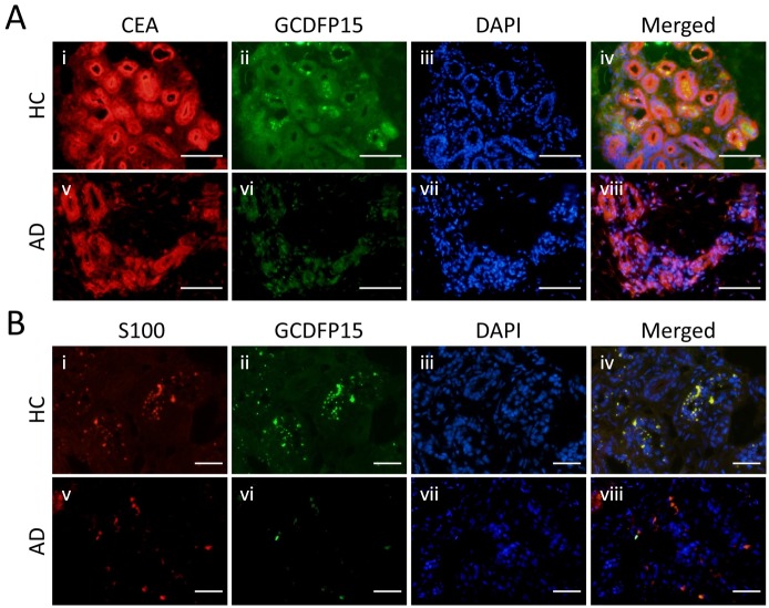
Fig 3. Distribution patterns of GCDFP15, CEA, and S100 protein in eccrine gland secretory coil.
(A) Double staining of CEA and GCDFP15 in the eccrine gland secretory coil. The expression of CEA is observed in whole eccrine gland secretory coil cells. (B) Double staining of S100 protein and GCDFP15. The expression of S100 protein is observed in a part of eccrine gland secretory coil cells as granular pattern. Colocalization of S100 protein and GCDFP15 is shown in yellow in the merged image. Bars indicate 50 μm.

