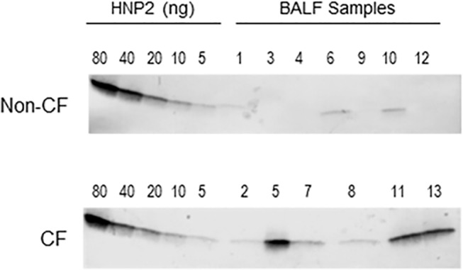
Fig 3. Quantification of HNP1-3 by Western immunoblot.

HNP2 peptide standard and bronchoalveolar lavage fluid (BALF) samples were separated by 4–20% SDS PAGE, blotted onto PVDF-PSQ membranes, probed with polyclonal rabbit antibodies against HNP1-3, and antibody binding was visualized with goat-anti rabbit antibodies conjugated to alkaline phosphatase and NBT/BCIP substrate. HNP1-3 differ by one amino acid only and co-migrate in this gel system. Per lane, the equivalent of the following BALF volumes were loaded: for Non-CF samples, 1: 20 μL, 3: 40 μL, 4: 40 μL, 6: 20 μL, 9: 40 μL, 10: 20 μL, 12: 20 μL; for CF samples, 2: 6 μL, 5: 6 μL, 7: 6 μL, 8: 20 μL, 11: 6 μL, 13: 6 μL.
