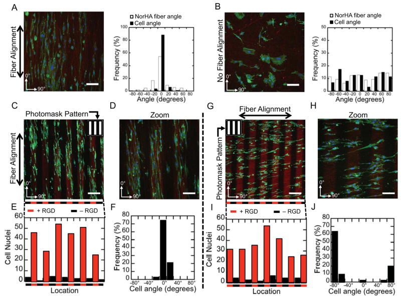
Figure 4.
Altered cell behavior in the presence of topographical cues and spatial patterning of RGD. (A) Cells orient and elongate with aligned nanofibrous topography on NorHA scaffolds with uniform RGD, (B) whereas cell orientation and elongation is abrogated in the absence of aligned nanofibers. Scale bars: 100 μm. (C–D) Cells orient and elongate with aligned nanofibrous topography on 100 μm wide cell adhesive lines of RGD parallel to nanofiber orientation. Scale bars: (C) 200 μm, (D) 100 μm. (E) Counts of nuclei (DAPI) as a function of horizontal position, and (F) angle of cells from image (C). (G–H) Cells elongate and orient with aligned nanofibrous topography (horizontal) but are spatially restricted (vertically) when RGD is patterned in 100 μm lines perpendicular to nanofiber orientation. Scale bars: (G) 200 μm, (H) 100 μm. (I) Counts of nuclei (DAPI) as a function of horizontal position, and (J) angle of cells from image (G). Staining in all images: Green: F-Actin (FITC-Phalloidin), Blue: Nuclei (DAPI), Red: RGD (GCDD-Rho).

