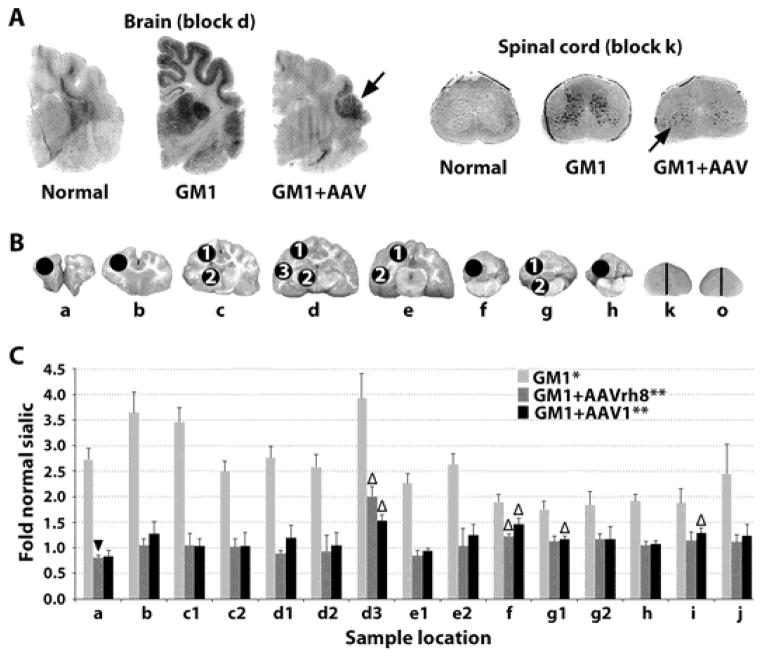
Fig. 2. Storage in the CNS of GM1 cats 16 weeks post-treatment.
(A) Storage in untreated GM1 cats was visualized by dark PAS staining in the gray matter and thalamus. In treated brains, residual ganglioside storage was present in focal areas (black arrows) of the temporal lobe (block d in panel B) and cervical spinal cord (block k in panel B). (B) Sample sites for sialic acid quantitation (circles) in brain (a–h) and spinal cord (k and o correspond to Fig. 1A; half of each block was used). (C) Sialic acid levels were measured in untreated GM1 cats (n = 4) and after treatment with AAVrh8 (n = 4) or AAV1 (n = 3) treated GM1 cats for comparison to normal healthy cats (n = 4). *, all samples from untreated GM1 cats were significantly higher than normal (P ≤ 0.015 for each block); **, all samples from treated GM1 cats were significantly lower than untreated GM1 cats (P ≤ 0.026 for each block) except AAV1 block h, as only 2 samples were available (P = 0.053); △, indicates samples from treated GM1 cats that were significantly higher than normal (P ≤ 0.035); ▼, indicates a sample from treated GM1 cats that was significantly lower than normal (P = 0.033). The Wilcoxon signed rank test was used for statistical comparisons.

