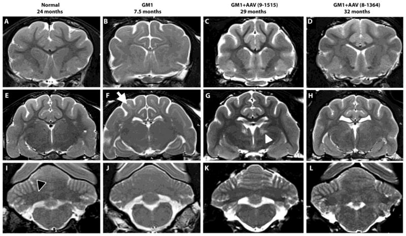
Fig. 5. MRI evaluation of GM1 cats.
T2-weighted MRI images (3 Tesla) were taken at the level of the caudate nucleus (A–D), thalamus (E–H) and deep cerebellar nuclei (I–L). Cortical white matter is hypointense to (i.e., darker than) gray matter in normal healthy cats, but hyperintense to (i.e., lighter than) gray matter in untreated GM1 cats (white arrow in panel F). Also, the deep cerebellar nuclei area is hypointense to surrounding gray matter in normal healthy cats (outlined black arrowhead in panel I), but becomes isointense with disease progression in untreated GM1 cats. In AAV-treated GM1 cats (GM1+AAV), hypointensity of cortical white to gray matter and deep cerebellar nuclei area to cerebellar gray matter was largely preserved, indicating reduced myelin loss after treatment. Both AAV-treated cats were clinically normal at the time of imaging. In 5 of 11 treated cats tested, a locus of hyperintensity was noted in the thalamus (white arrowhead in panel G), though no clinical or histopathological correlates have been identified to date. Ages in months are shown for each cat.

