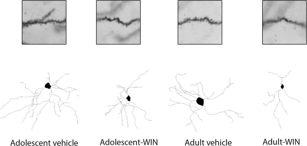
Fig. 4.
Representative spine micrographs and camera lucida drawings of nucleus accumbens (Acb) neuron following chronic WIN 55,212-2 administration in adolescent and adult Sprague–Dawley male rats. Included in the analysis are the core and shell subregions of Acb. Core subregion surrounds the anterior commissure, while the shell subregion is located medial and ventral to the lateral ventricle

