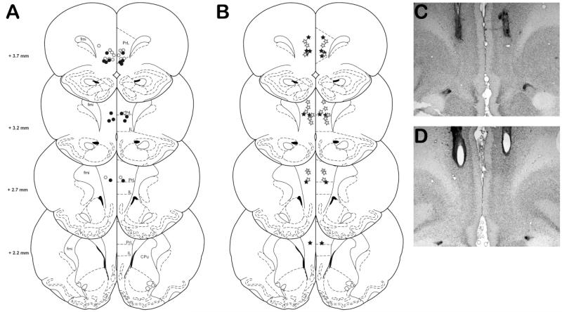
Fig 1.
Location of injector tips for rats tested in all versions of the rGT. A) Location of injector tips for rats tested with either BMI (filled circles, n=8) or LAG (open circles) in the standard rGT task. B) Location of injector tips for rats tested in the punishment (filled stars, n=8) and reward (open stars, n=12) versions of the rGT. C and D) Representative photomicrographs of cannulua placements within the prefrontal cortex. Rats with cannula placements outside of the PrL and IL were excluded from analysis (not shown). fmi (forceps minor corpus callosum); PrL (prelimbic cortex), IL (infralimbic cortex), CPu (caudate putamen). Adapted from Paxinos and Watson (2009).

