
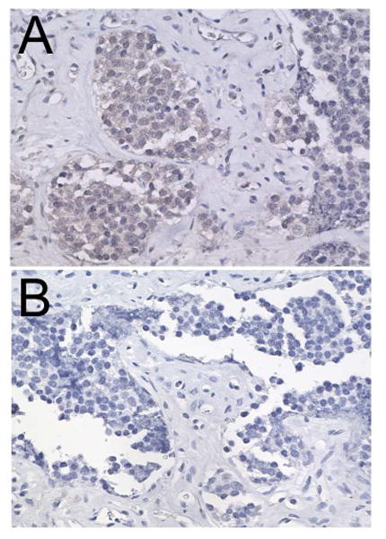
Figure 3.
Photomicrograph of immunohistoperoxidase staining for tyrosine hydroxylase showing faint immunoreactivity in the cytoplasm (A) compared to a control section lacking primary antibody (B).
Official websites use .gov
A
.gov website belongs to an official
government organization in the United States.
Secure .gov websites use HTTPS
A lock (
) or https:// means you've safely
connected to the .gov website. Share sensitive
information only on official, secure websites.
Photomicrograph of immunohistoperoxidase staining for tyrosine hydroxylase showing faint immunoreactivity in the cytoplasm (A) compared to a control section lacking primary antibody (B).