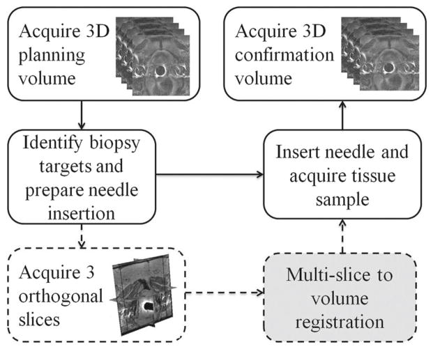
Fig. 1.
Clinical workflow of the image acquisitions during MRI-guided prostate biopsy. The solid lines indicate the standard biopsy procedure, and the dashed lines represent the additional orthogonal image slice acquisition specific to our study. The gray box is the motion compensation registration method we propose to incorporate

