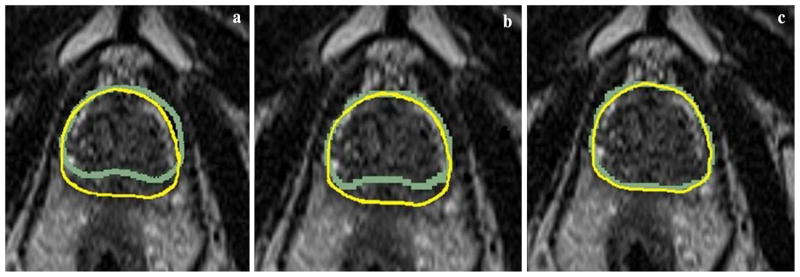
Fig. 4.

Examples of clinical image prostate contour overlay in the transverse plane. Each of the three images is copies of the same fixed image overlaid with the contours from the moving image a before registration, b after rigid registration, and c after deformable registration
