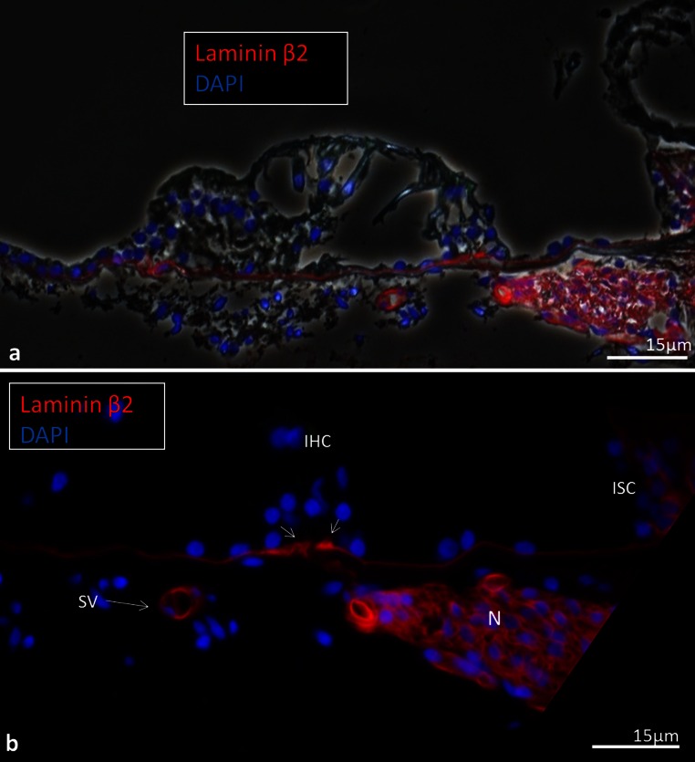
Fig. 12.
Laminin-β2 immunohistochemistry of the human organ of Corti. a Combined bright-field and fluorescent microscopy of the human organ of Corti and lamina spiralis with associated supplying nerves. b Fluorescent microscopy of the epithelial basement membrane. The thickness of the basement membrane increases in the habenular region (small arrows). Bar 20 μm. A spiral vessel shows strong laminin expression. There is no laminin staining in the organ of Corti. Hair and supporting cells are well preserved while the tectorial membrane is distorted. Bar 15 μm

