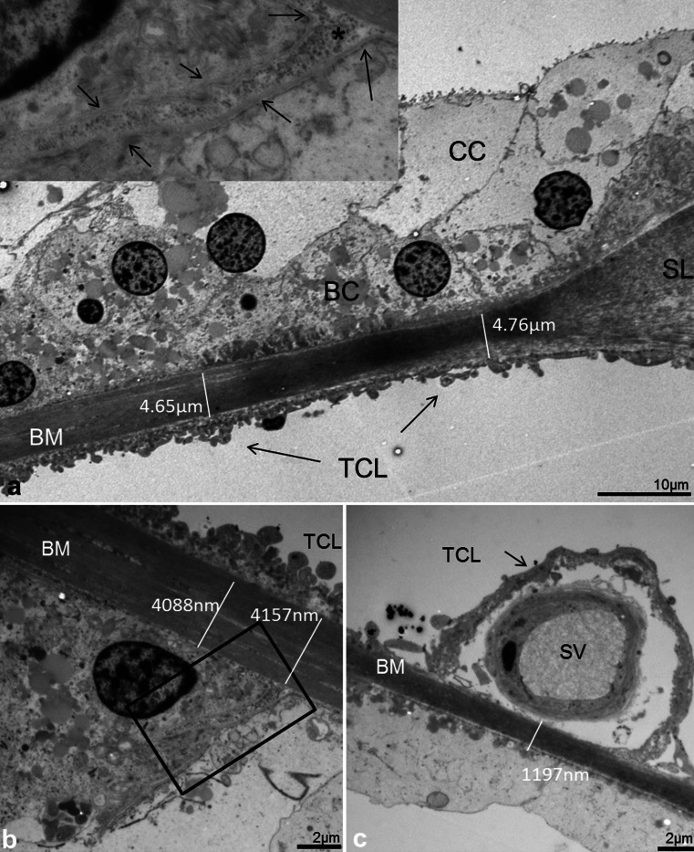
Fig. 6.
Thickness variation of the human BM in the hook (high-frequency) region portrayed with TEM. a Radial section shows the alternate radial thickness of the BM at the “hook” region. Laterally, the organ of Corti contains both Claudius (CC) and Boettcher cells (BC). The BM is over 4 μm thick and widens at the spiral ligament (SL). The electron-density of the BM is partly reduced and gives the impression that the BM is thinner near the SL. The tympanic covering layer (TCL) is thin. b BM thickness is slightly reduced medially beneath a Boettcher cell. An invagination of the basement membrane is seen between a Boettcher and Hensen cell (arrows, framed area is magnified in inset in a). c BM thickness is additionally reduced medially beneath the pillar feet at the spiral vessel (SV)

