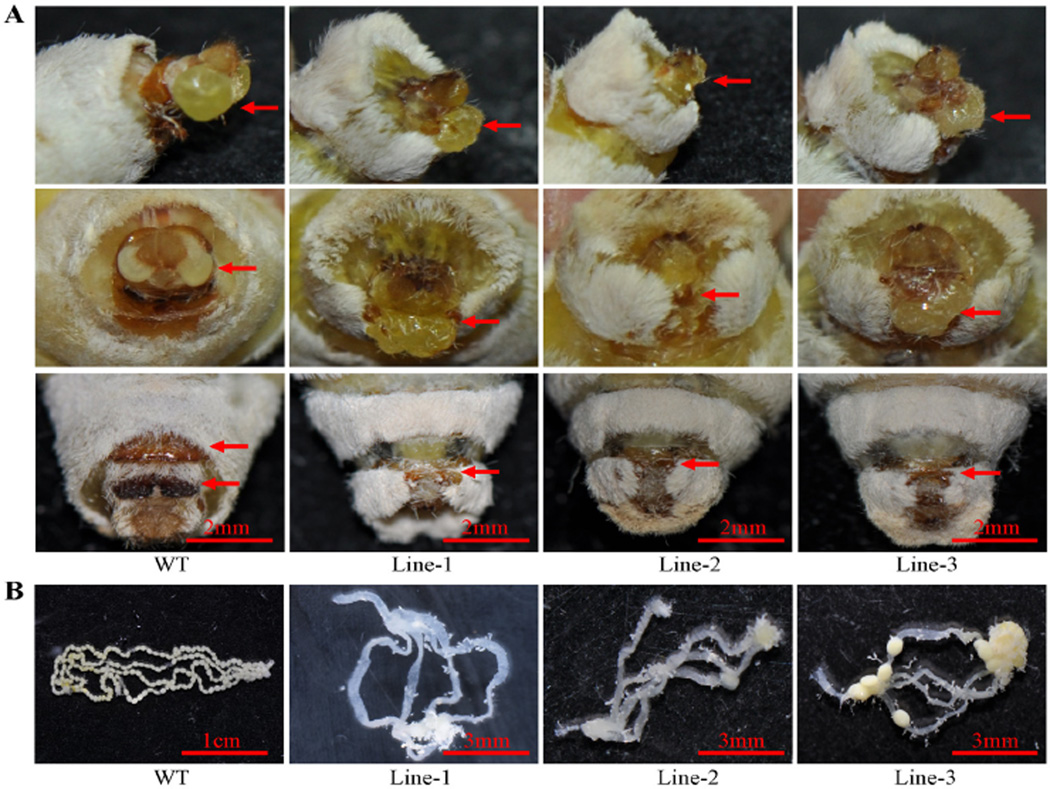
Figure 3.
Photographs of external genitalia and ovipositors of mutant females. (A) External genitalia of mutant females: lateral view (upper panel) and front view (middle panel) show the genital morphology of wild-type and mutant line 1–3 individuals. Red arrows indicate morphology of genital papilla. The lower panel shows that the female chitin plate is absent in the mutant individuals. Red arrows indicate morphology of chitin plate. (B) Mutant females have no (lines -1 and -2) or few (line-3) eggs in their ovarioles.

