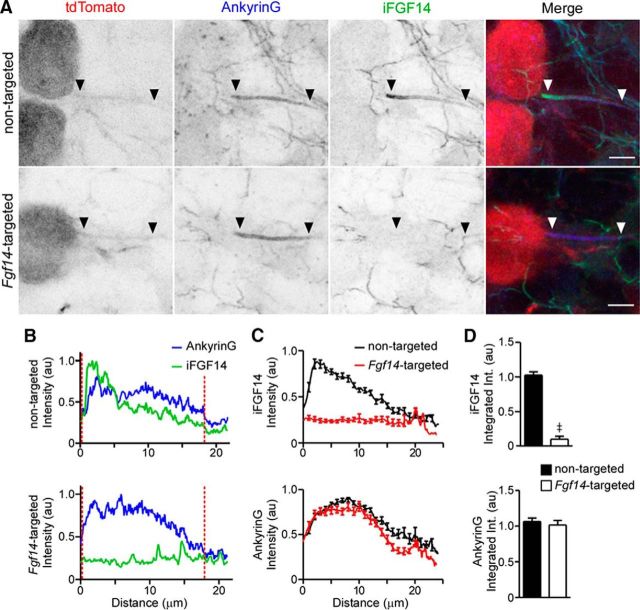
Figure 2.
Ankyrin G expression at the axon initial segment is robust in Purkinje neurons transduced with the Fgf14-targeted shRNA. A, Representative images of nontargeted and Fgf14-targeted shRNA-transduced Purkinje neurons identified by tdTomato fluorescence (red) and stained with anti-Ankyrin G (blue) and anti-iFGF14 (green) specific antibodies. In each panel, the arrowheads indicate the AIS. Scale bars, 5 μm. B, Representative line scans of anti-Ankyrin G and anti-iFGF14 immunofluorescence intensities along the AIS of a nontargeted (top) and an Fgf14-targeted (bottom) shRNA-transduced Purkinje neuron. Vertical (red) dotted lines indicate the start and end of each AIS. C, Mean ± SEM anti-iFGF14 (top) and anti-Ankyrin G (bottom) immunofluorescence intensities along the AIS of Purkinje neurons transduced with either the nontargeted (n = 29 AIS, 2 animals) or the Fgf14-targeted (n = 26 AIS, 2 animals) shRNA. D, Mean ± SEM anti-iFGF14 (top) integrated immunofluorescence intensity was reduced significantly (≠p < 0.0001; Student's t test) in Fgf14-targeted shRNA-expressing (n = 26) compared with nontargeted shRNA-expressing (n = 29) Purkinje neurons, whereas mean ± SEM anti-ankyrin G integrated immunofluorescence intensities were not measurably affected.

