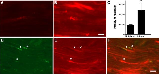
Figure 2.
L5SNT induces breakdown of BNB. A, B, Immunofluorescent micrographs showing that significantly more labeled GT1b–2b mAb (DyLight 594; red) deposits in the injured side sciatic nerve (B) compared with control side nerve (A) on postsurgical day 7. Scale bar, 10 μm. C, Quantitative analysis of labeled GT1b–2b deposition in uninjured vs injured nerve. n = 3. *p < 0.05. D–F, The injured sciatic nerve was immunostained for paranodal marker Caspr (green) and labeled GT1b–2b (red) binds to axon (*), node of Ranvier (arrowhead), and paranodal axon (arrow). Scale bar, 20 μm.

