
Figure 5.
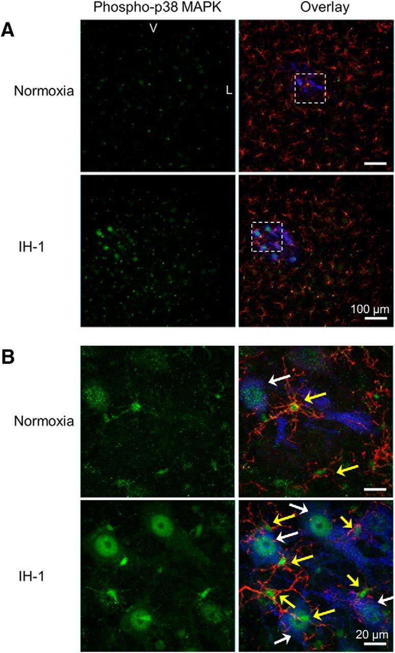
Phospho-p38 MAPK immunofluorescence is prevalent in back-labeled phrenic motoneurons and microglia of the ventral cervical spinal cord after IH-1. A, Confocal images (20×) show representative phospho-p38 MAPK (green) staining in the ventral cervical spinal cord after IH-1 (n = 6), which colocalized with back-labeled phrenic motoneurons (cholera toxin B (CtB); blue) and CD11b (microglia label; red). Minimal staining was evident after the normoxia treatment (n = 6, bottom). B, Higher magnification (100×) from the boxed area in A of the phrenic motor nucleus clearly shows colocalization with phrenic motoneurons and microglia after IH-1 (bottom). Less colocalization is evident after normoxia and is highlighted by white arrows (identifying CtB and phospho-p38 MAPK labeling) and yellow arrows (identifying CD11b and phospho-p38 MAPK labeling; top). V, Ventral; L, lateral.