
Fig. 1.
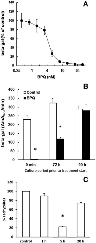
Inhibition of N. caninum proliferation by buparvaquone. (A) HFF were grown to confluence in a 96-well plate, treated with buparvaquone (BPQ) at various concentrations or DMSO as a solvent control and infected with N. caninum tachyzoites (103 per well) expressing the Escherichia coli beta-galactosidase gene. After 3 days, beta-galactosidase activity resulting from intracellular tachyzoites was determined. The activity is given as percentage of the solvent control. Mean values ± SE correspond to four replicates. (B) HFF were grown to confluence in a 96-well plate, infected with N. caninum tachyzoites (103 per well) expressing the E. coli beta-galactosidase gene. After different time points, the infected cells were treated with 100 nM buparvaquone (BPQ) or with DMSO as a solvent control. After 4 days, beta-galactosidase activity resulting from intracellular tachyzoites was determined. The activity is given as percentage of the solvent control. Mean values ± SE correspond to four replicates. The values marked by an asterisk are significantly different from the corresponding control value (two-sided t-test, P < 0.01). (C) HFF grown in 6 well plates were treated with 1 µM BPQ for 1 or 5 h, washed with PBS, and infected with 2 × 10⁵N. caninum tachyzoites/well. After 4 days, cells were harvested and subjected to quantitative real time PCR. Results are shown as % proliferation in relation to untreated controls. Mean values ± SE correspond to four replicates. The values marked by asterisks are significantly different from the corresponding control value (two-sided t-test, P < 0.01).