
Fig. 3.
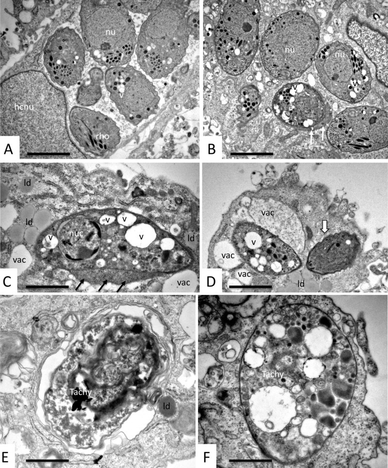
Longer-term BPQ treatment (24–120 h) of N. caninum tachyzoites cultured in human fibroblasts alters the ultrastructure of N. caninum tachyzoites. (A and B) Typical specimens fixed and processed after 72 h of drug treatment. Tachyzoites were found either in large parasitophorous vacuoles (A), or the parasitophorous vacuole was not discernible (B), and some tachyzoites had developed electron-dense accumulations at their periphery (white arrows in B). In any case, tachyzoites exhibited an increased cytoplasmic vacuolization (v). hcnu = host cell nucleus, nu = tachyzoite nucleus, rho = rhoptries. Ultrastructural changes were more evident after 120 h of BPQ treatment. In about 80% of cases parasites were still recognizable, but clearly damaged (C, D), with condensed nucleus (nuc), aberrant cytoplasmic organization with large vacuoles (v). There is no parasitophorous vacuole visible, but electron dense material accumulating at the tachyzoite periphery (black arrows in C), vacuolization in the host cell cytoplasm adjacent to the parasites (vac), and formation of many lipid droplets (ld). (E and F) Examples of completely distorted tachyzoite residues (Tachy), either packed into a vacuole and surrounded by several membranes (E), or as a single entity delineated by a still intact plasma membrane only (F). Bars in A and B = 1.5 µm, C = 1.7 µm, D = 1.6 µm, E and F = 0.9 µm.