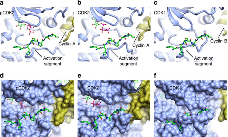
Figure 3. The CDK substrate binding site.
(a,d) T160-phosphorylated CDK2–cyclin A (1QMZ,50); (b,e) unphosphorylated CDK2–cyclin A (PDB code 1FIN,43); (c,f) unphosphorylated CDK1–cyclin B. Each complex in each row is shown in the same view, pCDK and CDK designate the phosphorylated and unphosphorylated proteins, respectively. CDK and cyclin subunits are coloured ice-blue and gold, respectively. In all panels the peptide from 1QMZ is shown as a reference for the site of peptide substrate binding and is drawn in cylinder mode with carbon atoms coloured green. (a–c) Ribbon representation of the proteins. (d–f) Molecular surfaces of the proteins to highlight shape complementarity and/or steric clashes.

